


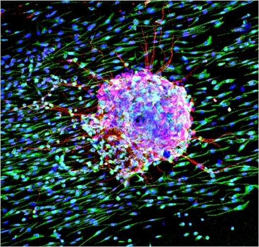
Direct Laser Fabrication of Biomimetic, 3D Scaffolds for Tissue Regeneration
Direct Laser Fabrication of Biomimetic, 3D Scaffolds for Tissue Regeneration
Short Description and Main Findings of the Research Topics:
Research Topic 1: Direct Laser Micro/Nano Fabrication of Biomimetic Scaffolds
Short description
The aim is to investigate the biocompatibility of laser-engineered biomimetic 3D scaffolds fabricated on hard metallic and soft polymeric materials, exhibiting different micro/nano topographies and surface energies.
Abstract
The extracellular matrix provides the necessary cues at micro and nano-scale for cell adhesion, alignment, proliferation and differentiation. In this context, the surface topography of biomaterials can have an important impact on cellular adhesion, growth and proliferation. Apart from the overall roughness, the detailed morphological features, at all length scales, significantly affect the cell-biomaterial interactions in a plethora of applications including structural implants, tissue engineering scaffolds and biosensors.
The main objective is to investigate the biocompatibility of laser-engineered biomimetic 3D scaffolds fabricated on hard metallic and soft polymeric materials, exhibiting different micro/nano topographies and surface energies. Ultrafast pulsed laser irradiation is considered as a simple, precise and effective microfabrication method to produce structures of controlled geometry and pattern regularity. The variation of irradiation parameters, such as fluence and irradiation environment gives rise to significant changes of the surface morphology attained (i.e. geometry, dimensions and density of the structures). As a consequence, morphologies ranging from microcones to nanoripples (Figure 1), as well as hierarchical micro/nano structures (Figure 2) can be fabricated and further used as cell culture platforms.
The laser fabricated scaffolds with controlled surface roughness, wettability and surface energy can be used as model platforms to study the influence of topography on cell response. It is demonstrated that, depending on the laser processing conditions, distinct cell-philic or cell-repellant patterned areas can be attained with a desired motif (Figure 3). Laser processing could thus enable spatial patterning of cells in a controllable manner, giving rise to advanced capabilities in cell biology research.
Research has shown that cell adhesion and migration could be tuned via the laser-patterned substrates. It was also shown that microconical substrates could influence sympathetic and sensory neuronal alignment as well as NGF-induced PC12 cell differentiation (see also Research Topic 2).
Scaffolds on hard materials
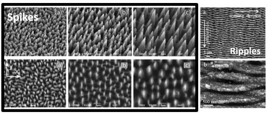
Figure 1: Various types of femtosecond laser fabricated scaffolds on hard materials with feature sizes ranging from a few hundreds of nanometers to tens of microns (C Simitzi, P Efstathopoulos, A Kourgiantaki, A Ranella, I Charalampopoulos, C Fotakis, I Athanassakis, E Stratakis, A Gravanis, Biomaterials, 2015, 67: 115-128, doi.org/10.1016/j.biomaterials.2015.07.008).
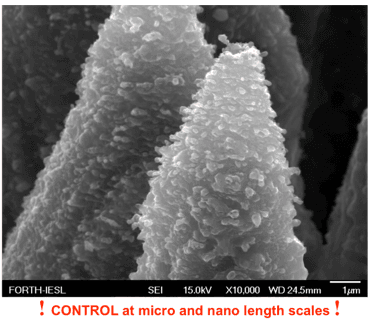
Figure 2: Laser fabricated arrays of biomimetic hierarchical micro/nano conical structures(Chara Simitzi, Pascal Harimech, Syrago Spanou, Christina Lanara, Amelie Heuer-Jungemann, Aleka Manousaki, Costas Fotakis, Anthi Ranella, Antonios G Kanaras, Emmanuel Stratakis, Biomater. Sci., 2018,6,1469, doi: 10.1039/c7bm00904f)
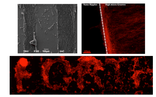
Figure 3: Patterning of Schwann (SW10) cells cultured on laser fabricated substrates exhibiting cell-philic and cell-repellant areas (Ch Yiannakou, Ch Simitzi, A Manousaki, C Fotakis, A Ranella, E Stratakis, 2017 Biofabrication 9 025024, https://doi.org/10.1088/1758-5090/aa71c6)
Scaffolds on soft materials
a) Replicated Scaffolds
Soft lithography has been successfully used to transfer well-defined micro-sized patterns from hard materials to soft (bio)materials. The replication of micro/nano topographies is realised on polymeric systems, such as poly (lactic-co-glycolic acid) (PLGA), polycaprolactone (PCL) (Figure 4) and polydimethylsiloxane (PDMS), in order to investigate the effect of material surface energy and stiffness on cellular responses (adhesion, proliferation, survival, growth and differentiation).
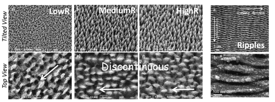
Figure 4a: SEM images (tilted view) of PLGA replicas with the hierarchical (microcone-spikes and nano-ripples) topographies (Babaliari, E., Kavatzikidou, P., Angelaki, D., Chaniotaki, L., Manousaki, A., Siakouli-Galanopoulou, A., Ranella, A., & Stratakis, E. (2018). Engineering Cell Adhesion and Orientation via Ultrafast Laser Fabricated Microstructured Substrates. International journal of molecular sciences, 19(7), 2053, doi: 10.3390/ijms19072053).
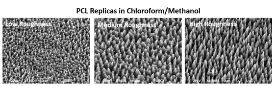
Figure 4b: SEM images (tilted view) of PCL replicas exhibiting various microcone topographies.
b) 3D scaffolds of porous collagen via subtractive laser manufacturing:
Laser micromachining provides a novel CAD/CAM rapid prototyping microfabrication process that can provide complex implant designs based on an established biomaterial utilized in clinical practice. It is shown that fs laser micromachining of porous collagen (Figure 5), in particular, can fabricate high-precision micron-sized features (e.g. canals, wells) and provides novel ways to modulate the microenvironment felt by interacting cells, tailor implants to the needs of individual patients, or tools to meet the current major challenges of regenerative medicine.
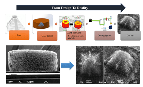
Figure 5: fs laser micromachining of porous collagen scaffolds
Research Topic 2: 3D Scaffolds Hosting Neurons and Neural Stem Cells
Short Description
The aim is to develop laser-engineered micro/nano scaffolds (3DLS) for hosting 3D cultures of neural stem cells.
Abstract
Neural stem cells (NSCs) are intrinsically capable of differentiating into different neural cell types: neurons, oligodendrocytes and astrocytes, and have emerged as important players in the generation and maintenance of neural tissue as well as in treating neurodegenerative diseases and neurological injuries. However, successful development of NSC-based therapies requires more sophisticated technologies from the ones that are already available and deeper understanding of NSCs’ functions. The NSCs reside in a complex three-dimensional (3D) niche in vivo where they are exposed to a plethora of signals, including physical signals such as tensile, compressive and shear stresses, discontinuities and differences in roughness of the ECM molecules. Topography is capable of inducing different effects on NSCs, such as changes in cell morphology, alignment (Figures 1 and 2), adhesion, migration, proliferation, cytoskeleton organization and also differentiation (Figure 3). However, simulating this 3D environment for NSC culture and subsequent development of 3D neuronal networks that maintain functional neuronal properties (synaptogenesis and neurotrophic performance) remains a challenge.
To respond to this challenge we have fabricated 3D laser-engineered micro/nano scaffolds (3DLS) featuring different micro/nano topographies for hosting neurons, glia and NSCs. These are advantageous platforms to study the biology of NSC proliferation, differentiation, neuritogenesis and synaptogenesis. Patterning of neuronal outgrowth in vitro is important in tissue engineering as well as for the development of neuronal interfaces with desirable characteristics. Laser-patterned biomimetic scaffolds could potentially be a useful platform for patterning neurons into artificial networks, allowing the study of neuronal cells interactions under 3D ex-vivo conditions.
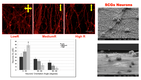
Figure 1: SCGs neurons orientation on laser fabricated discontinuous anisotropic microconical substrates (C Simitzi, P Efstathopoulos, A Kourgiantaki, A Ranella, I Charalampopoulos, C Fotakis, I Athanassakis, E Stratakis, A Gravanis, Biomaterials, 2015, 67: 115-128, doi.org/10.1016/j.biomaterials.2015.07.008).
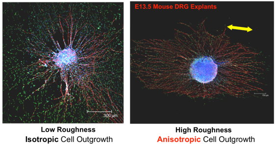
Figure 2: DRG cultures on laser fabricated discontinuous anisotropic microconical substrates (C Simitzi, P Efstathopoulos, A Kourgiantaki, A Ranella, I Charalampopoulos, C Fotakis, I Athanassakis, E Stratakis, A Gravanis, Biomaterials, 2015, 67: 115-128, doi.org/10.1016/j.biomaterials.2015.07.008).
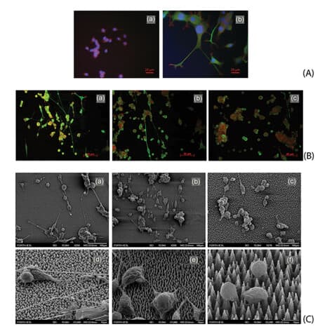
Figure 3: Effect of surface roughness on PC12 cell differentiation. (C. Simitzi, E. Stratakis, C. Fotakis, I. Athanassakis and A. Ranella, Microconical silicon structures influence NGF-induced PC12 cell morphology, J Tissue Eng Regen Med2015;9: 424–434, DOI:10.1002/term.1853)
Research Topic 3: Development of microfluidic systems for cell studies under dynamic culture conditions
Understanding the cell-biomaterial interaction under dynamic culture conditions, in vitro, is potentially useful in the fields of tissue engineering and regenerative medicine.
A precise flow controlled microfluidic system with specific custom-designed chambers, incorporating laser-microstructured polyethylene terephthalate (PET) substrates comprising microgrooves, is developed to assess the combined effect of shear stress and topography on cells’ behavior (Fig.1). Specifically the dynamic cultures are performed for the study of the cytoskeleton, directionality and proliferation of cells on micro-nano patterns. A comparison between static and dynamic cultures is always performed in combination with computational flow simulations to calculate accurately the shear stress values. The main findings demonstrate that wall shear stress gradients may be acting either synergistic or antagonistic depending on the substrates groove orientation relative to the flow direction (Fig.2, Fig.3).
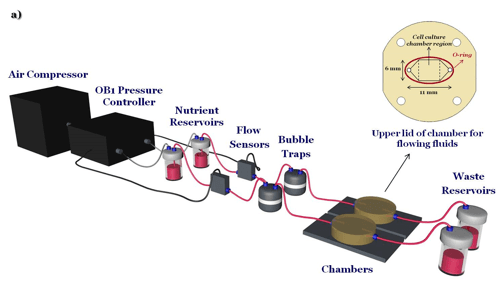
Figure 1: Custom-designed Microfluidic System
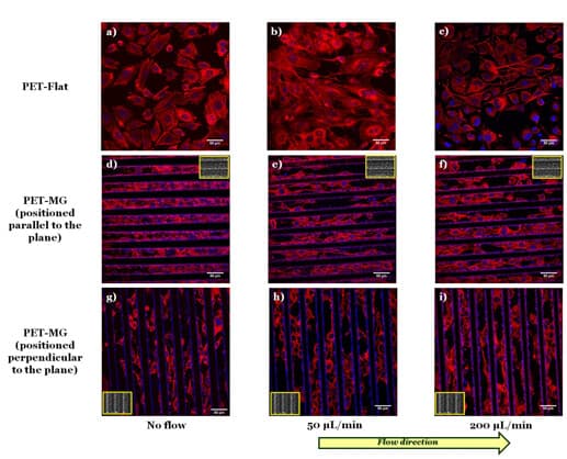
Figure 2: Confocal images of SW10 cells cultured on the PET-Flat (a, b, c) or inside the MG of the PET-MG substrates (d-i), under static (a, d, g) or dynamic conditions, applying 200 (c, f, i) μL/min, on the third day of culture. The cytoskeleton of the cells is visualized with red color (Alexa Fluor® 680 Phalloidin) while the nuclei with blue color (DAPI). The direction of the flow was parallel (f) or perpendicular (i) to the microgrooves. The inset SEM images, framed by a yellow box, depict the geometry of microgrooves (paper under submission).
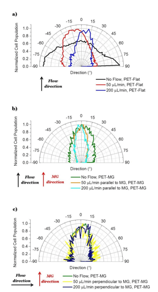
Figure 3: Directional polar plots of cells’ cytoskeleton: a) No flow, PET-Flat (black line) 200 μL/min, PET-Flat (blue line), b) No flow, PET-MG (green line) – 200 μL/min parallel to MG, PET-MG (turquoise line), c) No flow, PET-MG (green line) - 200 μL/min perpendicular to MG, PET-MG (dark blue line). The black and red arrows represent the direction of the flow and the microgrooves, respectively.
Moreover, an additional microfluidic chamber incorporating polymeric microgrooved substrates that could be inserted to a non-linear microscope for live imaging observation is developed for nanotoxicology studies. The effect of nanoparticles on cells is examined in terms of survival and proliferation both under static and dynamic conditions (Fig.4).
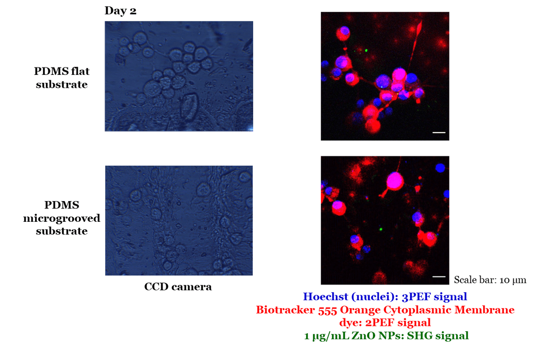
Figure 4: Observation of live NIH 3T3 cells cultured on PDMS flat and microgrooved substrates with ZnO NPs inside the microfluidic chamber, under static conditions, with the NLO microscope. Scale bar: 10 μm.
In collaboration with Mary Kefalogianni and Dr. Sotiris Psilodimitrakopoulos
Main research expertise of the LaBio team:
•Direct laser fabrication of hard and soft biomaterials customized, personalized and on-demand for more than one application in the Medical field (Circular Economy Action Plan)
•Soft lithography for replicating the laser micro-nano structures/patterns on bio-based biodegradable polymers (e.g. PLGA, PCL) with substantially lower environmental footprint
•3D & 4D printing (Optimisation of Additive manufacturing technique) as part of the global vision of sustainability (4D Printing Subgroup)
•Use of renewable materials for 4D printing, after the growing phenomenon of plastic waste worldwide eg various natural materials (collagen, cellulose) and synthetic (PLA)
•In-vitro degradation studies of the bio-based biodegradable polymers
•Characterisation techniques for the study of their physical, morphological, optical and chemical properties
•In-vitro cytotoxicity testing of the developed patterned surfaces
•In-vitro cytocompatibility studies of various cells (eg. Mouse neuronal cell lines, mouse neural stem cells, mouse mesenchymal stem cells, fibroblasts) on the above biomaterial architectures/patterns (in collaboration with TERMIM lab)
•Proteomic and biomolecular profiles of cell lines adhered on patterned bio-based surfaces (in collaboration with TERMIM lab)
•Study of the effect of shear stress on cells adhered on polymeric patterned surfaces (in collaboration with TERMIM lab)
•Ca+2 imaging, electrical activity and live imaging of neural stem cells and neuronal cell lines interacting with bio-based materials via a non-linear microscopy system (in collaboration with the Live Cell Imaging Subgroup)
•Bioactive scaffolds for local drug delivery applications
•Biocompatible Nanoparticles (in collaboration with the Nanomaterials Subgroup )
•Live imaging and cytotoxicity of nanoparticles under static and dynamic conditions (in collaboration with the Live Cell Imaging Subgroup)
Main medical applications: neural implants, bone implants, corneal implants, drug delivery etc